An 80 year old male is bought to your ED via ambulance following a syncopal episode. He reports sitting on a church pew, when he apparently collapsed without prior warning. According to bystanders he was unresponsive on the ground looking pale then ‘blue’. He was making some respiratory effort and eventually recovered without intervention.
By the time you examine him, he is alert and oriented (though, amnestic to the actual event). His pulse is 60, he is warm and perfused (with a BP of 138/66). There is no evidence of cardiac failure and his neurological exam is unremarkable. You do note a pacemaker box in his upper left chest and his CXR shows that this is a ‘dual-lead’ variety….
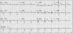
This is his ECG.
What’s going on here ?
How do you explain his syncope ??
What needs to happen now ???
- Atrial paced rhythm @ 60/min
- Left Axis Deviation
- PR 280-300msec. QRS 150msec (RBBB pattern). QTc ~460msec
- Left Anterior Fascicular Block (LAD, ‘rQ in inferior leads)
- No obvious pronounced ischaemia.
Impression.
- Atrial paced rhythm
- Trifasicular block.
?Syncope secondary to complete heart block (whilst atrially paced)
– why didn’t his ventricular pacing kick in ??
– is there a pacemaker malfunction ??
??tachydysrhythmia
He is admitted to Coronary Care for telemetry whilst he awaits a pace-maker interrogation…..
Pacemakers.
In short, this case prompted some further reading on PPMs & AICDs, particularly the codings, the variety of settings available & how each functions to assist native cardiac activity (or lack thereof…..).
What’s in one … ??
- Pulse generator (& battery), electronic circuitry and leads.
- Lithium power (lasting 4 to >10 years)
- Leads are placed into ventricular (+/- atrial) endocardium
- Can be unipolar or bipolar
- Bipolar leads are compatible w/ AICD systems, but are larger & more prone to fracturing.
What does it do … ??
- Two basic functions:
- stimulate the heart electrically.
- sense the intrinsic cardiac activity (native rhythm)
- Additional functions:
- Overdrive pacing
- Deliverance of shock/defibrillation
Who gets it … ??
- 3rd-degree & advanced 2nd degree AV block w/
- symptomatic bradycardia (incl. heart failure) or ventricular dysrhythmia
- symptomatic bradycardia (secondary to drugs required for dysrhythmia therapy)
- Documented asystole (>3sec) or escape rhythm (<40bpm) or escape rhythm (below AV node) in an asymptomatic individual
- After catheter ablation of the AV node, or if post-operative AV blockade is unlikely to resolve.
- Neuromuscular disease w/ AV block (eg. muscular dystrophy)
- Symptomatic bradycardia from 2nd-degree AV block
- Asymptomatic, persistent 3rd-degree block
- Chronic bifascicular or trifascicular block w/ intermittent 3rd-degree block (or Type II 2nd-degree block)
- 2nd or 3rd degree block w/ exercise
What do those codes mean … ??
Examples:
- AAI:
- Atrial pacing and sensing.
- Intrinsic atrial activity inhibits pacemaker firing & natural/native conduction occurs.
- Lack of intrinsic activity (beyond a programmed time frame) triggers an atrial paced beat (which results in normal AV conduction and ventricular contraction).
- VDD:
- Capable of pacing ventricle only.
- Senses both atrial & ventricular native depolarisation
- If intrinsic ventricular depolarisation occurs; responds by dual inhibition of both atrial & ventricular pacing.
- A paced ventricular beat is triggered in response to a sensed intrinsic atrial depolarisation.
- VVI:
- Ventricular (only) pacing and sensing.
- A paced ventricular beat will be triggered if an intrinsic depolarisation fails to occur during a set, programmed interval.
- A native ventricular beat will inhibit pacemaker firing.
- DDD:
- Can sense & pace both atrium and ventricle.
- Atrial pacing initiated if native atrial contraction fails to occur in given time frame.
- Ventricle is paced if native ventricular contraction fails to occur for a given time frame following atrial activity.
- Native sinus rhythm inhibits both atrial and ventricular pacing.
What about the magnet … ??
Magnet application (placed externally over the pulse generator in the chest wall) causes closure of a reed-switch within the pace-maker circuitry, converting the pacemaker to an asynchronous or fixed-rate pacing mode (set by the manufacturer), and the pacemaker is no longer inhibited by the patient’s intrinsic electrical activity.
- Most commonly used when the patient’s intrinsic heart rate exceeds the pacemaker’s set rate and pacemaker function is inhibited.
the conclusion.
Well our fella’s pacemaker was interrogated a few hours later in the CCU.
- Turn’s out he was in an MVP-mode (technically a combination of AAI-DDD)
- Had had a few episodes of Wenchebach and two short runs of VT (‘weeks ago’).
- There was no dysrhythmic event at the time of his syncope.
- He was changed to DDD mode & discharged home 48 hours later with a presumptive diagnosis of vasovagal syncope.
MVP mode (a Medtronic program) is designed to reduce the risk of AF in patients with permanent pacemakers.
- MVP = managed ventricular pacing
- MVP is atrial-based pacing (aimed to significantly reduce unnecessary right ventricular pacing.
- Primarily operates in AAI whilst providing a safety-net of a dual-chamber back-up mode, if required
- Why ??
- Risk of AF doubles w/ ventricular pacing (DDD-L or DDD-S) versus atrial pacing (AAI-R).
References.
- Rosenʼs Emergency Medicine. Concepts and Clinical Approach. 7th Edition
- Pacemaker Rhythms – Normal Patterns @ lifeinthefastlane.com
– This page is a fantastic resource for further reading on pacemakers (particular ECG interpretation). - Managed Ventricular Pacing – Medtronic.com
- Nielsen JC et al. A randomized comparison of atrial and dual-chamber pacing in 177 consecutive patients with sick sinus syndrome: echocardiographic and clinical outcome. J Am Cardiol. August 20, 2003;42(4):614-623.


Pingback: The LITFL Review 099 - Life in the Fast Lane medical education blog